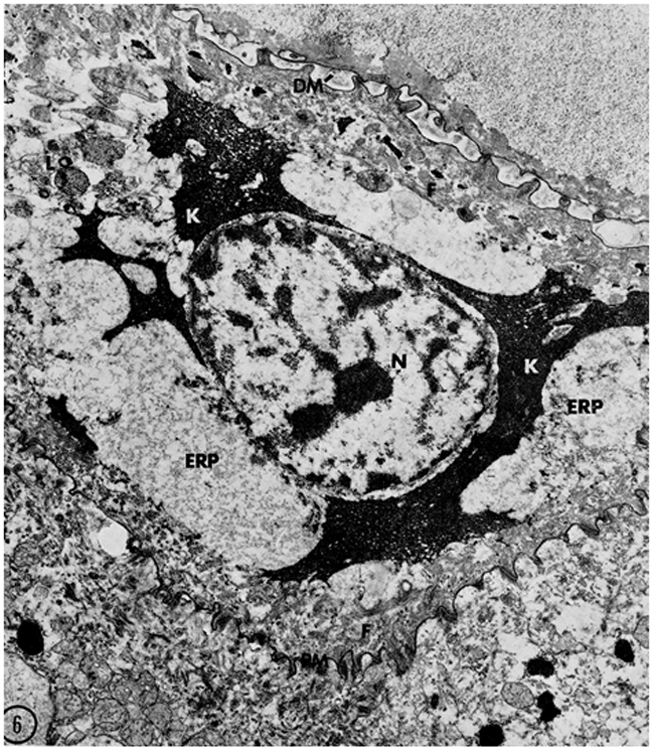
Figure 1.

A keratinocyte in an advanced stage of transformation from a granular cell to a corneocyte. Lysosomes/autophagosomes (L) are present in the cytoplasm. Filaggrin (K) is spread throughout the cytoplasm intermingling with keratin filaments (F). The thickened, modified corneocyte envelope (PM) is tightly attached to the lower granular cell. The nucleus (N), last of the recognizable organelles, shows signs of degradation. Reprinted from ©Lavker and Matoltsy, 1970, J Cell Biology, 44:501–512
