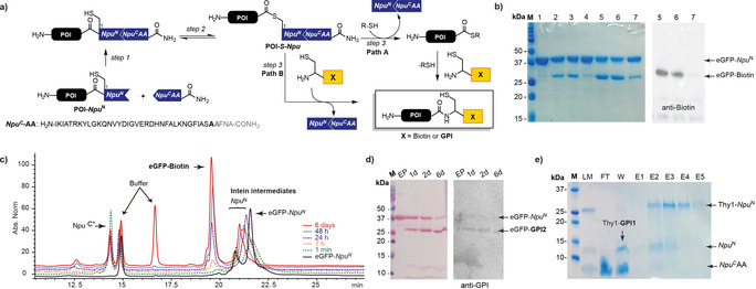
Figure 2.

One‐pot ligation of eGFP with biotin. a) Scheme of the OPL reaction. b) Comparison of OPL with MESNA, MMP, and MMBA by SDS‐PAGE and western blot (eGFP‐Biotin detected with anti‐biotin antibody). M=Molecular weight marker; 1) GFP‐Npu N; 2) OPL with MESNa, 1 h; 3) OPL with MMP, 1 h; 4) OPL with MMBA, 1 h; 5) OPL with MESNa, 1 d; 6) OPL with MMP, 1 d; 7) OPL with MMBA, 1 d. c) Kinetic study of OPL, monitored by RP‐HPLC (C4). d) SDS‐PAGE and western blot of the OPL of eGFP with GPI2; detection with Ponceau S staining and anti‐GPI antibody after 1,2 and 6 days. M=Molecular weight marker, EP=eGFP‐Npu N. e) SDS‐PAGE of OPL between Thy1 and GPI1; purification by His‐Trap. M=Marker; LM=ligation mixture 6 d; FT=flow‐through; W=wash; E=elution fractions. MESNa=sodium 2‐mercaptoethanesulfonate, MMP=methyl 3‐mercaptopropionate.
