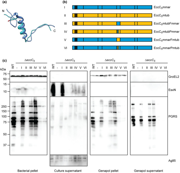
FIGURE 2.

Role of linker 2 in substrate specificity in M. marinum ESX‐5. (a) Superimposition of T. curvata linker 2 (bright blue) and linker 3 (dark blue). The 43 residues that were not resolved in the crystal structure are indicated in red. (b) Schematic overview of M. marinum and M. tuberculosis EccC5 as well as the chimeric constructs used to complement eccC5 mutants of these species. See Figure S4a for the exchanged sequences. (c) Secretion analysis of M. marinum ∆eccC5 complemented with WT eccC5mmar, eccC5mtub and chimeric constructs depicted in b. Proteins were visualized by SDS‐PAGE and immunoblotting using antibodies against EsxN and PE_PGRS proteins (ESX‐5 substrates), GroEL2 (lysis and whole‐cell loading control) and Ag85 (secreted fraction loading control)
