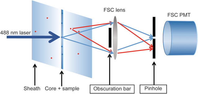
Figure 1.

Schematic representation of the optical configuration in front of the FSC detector. With a small pinhole in place, scatter light derived from a confined small area around the core stream only can reach the FSC detector (blue arrows), while light coming in with other angles (either stray light or scatter from occasional particles in the sheath fluid) are blocked (red arrows). The figure does not reflect actual relative sizes and dimensions. [Color figure can be viewed at wileyonlinelibrary.com]
