
Figure 1.
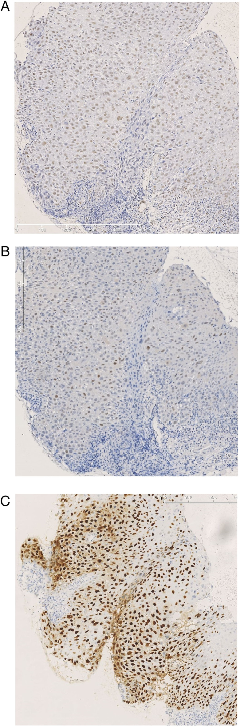
Example of a biopsy from a laryngeal tumor showing a high expression in immunohistochemical staining for (A) pATM, (B) pChk2, and (C) p53. Original magnification 200x. [Color figure can be viewed in the online issue, which is available at www.laryngoscope.com.]