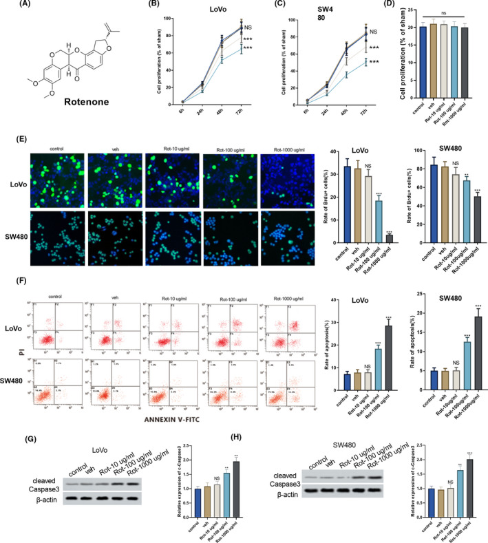
FIGURE 1.

Rotenone affected the proliferation and apoptosis of colon cancer cells. Colon cancer cells LoVo and SW480 were divided into control group, solvent control group (veh), and rotenone groups (Rot‐10 μg/mL, Rot‐100 μg/mL and Rot‐1000 μg/mL). A, Molecular structural formula of rotenone. B and C, The proliferation of LoVo and SW480 cells was detected using CCK8 assay. D, The proliferation of normal colon epithelial cell line FHC was detected using CCK8 assay. E, The proliferation of LoVo and SW480 cells was detected using Brdu staining. F, The apoptosis of LoVo and SW480 cells was detected using flow cytometry. G and H, Cleaved caspase 3 protein from LoVo and SW480 cells was detected using Western blot, **P < .01, ***P < .001 vs veh group. B and C,  , Rot‐1000 μg/mL;
  , Rot‐100 μg/mL;
  , Rot‐10 μg/mL;
  , veh;
  , control
