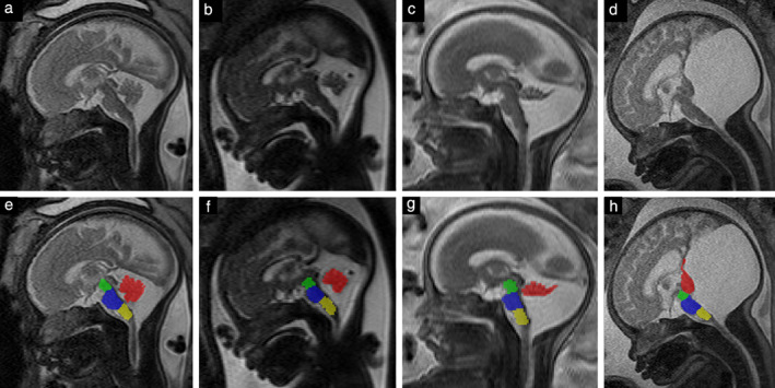
Figure 4.

Midsagittal T2‐weighted magnetic resonance images in fetuses with cystic posterior fossa malformation (a–d), and with corresponding segmentation of vermis (red), mesencephalon (green), pons (blue) and medulla oblongata (yellow) (e–h). (a,e) Group‐1 fetus at 32 + 4 weeks' gestation; (b,f) Group‐2 fetus at 29 + 2 weeks; (c,g) Group‐3 (Dandy–Walker malformation group) fetus at 25 + 4 weeks; (d,h) Group‐3 fetus (with more distinct findings) at 36 + 3 weeks.
