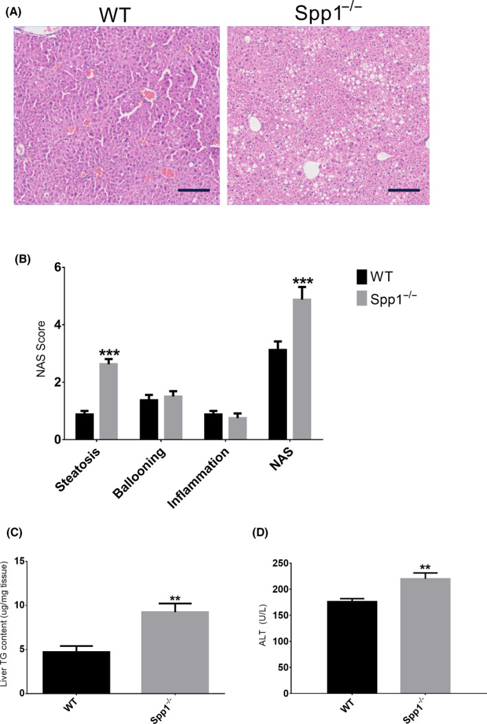
FIGURE 1.

Lack of osteopontin enhances hepatic steatosis in NASH/HCC mice. A, Representative H&E‐stained sections of WT (left panel) and OPN knock‐out (Spp1 −/−, right panel) NASH‐HCC livers. Scale bar = 100 µm. B, Non‐alcoholic fatty liver disease score (NAS), qualitatively evaluated out of the H&E‐stained sections. NAS is the sum of the scores attributed to the three hallmarks of NAFLD, ie steatosis, hepatocellular ballooning and inflammation. C‐D, Liver triacylglycerol content (C) and plasma alanine aminotransferase activity (D). **P ≤ .01, ***P ≤ .001. Tissues were harvested from 8‐week‐old mice (n = 8 per group). See also Figures S1 and S2
