FIGURE 4.

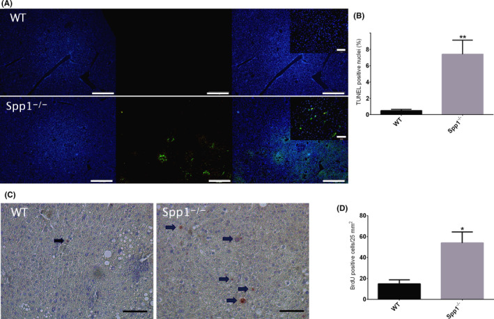
Enhanced apoptosis and proliferation in Spp1 −/− mice. A, TUNEL assay for the identification of apoptotic cells in WT (upper panel) and Spp1 −/− (lower panel) NASH‐HCC livers. Hoechst 33 342 (blue signal, left panels) stains all nuclei, while apoptotic cells are marked with green fluorescence (central panels). Right panels represent the merge of the two signals. Scale bar = 200 µm. Inserts at higher magnification, scale bar 50 µm. B, Graph of the proportion of TUNEL‐positive cells. C, BrdU staining for the identification of proliferating cells. Sections were incubated with anti‐BrdU antibody and counterstained with haematoxylin. Positive nuclei are depicted in red and highlighted by black arrows. Scale bar = 50 µm. *P ≤ .05, **P ≤ .01. D, Analysis of BrdU‐positive cells/25 mm2 area. Tissues were harvested from 12‐week‐old mice (n = 8 per group)
