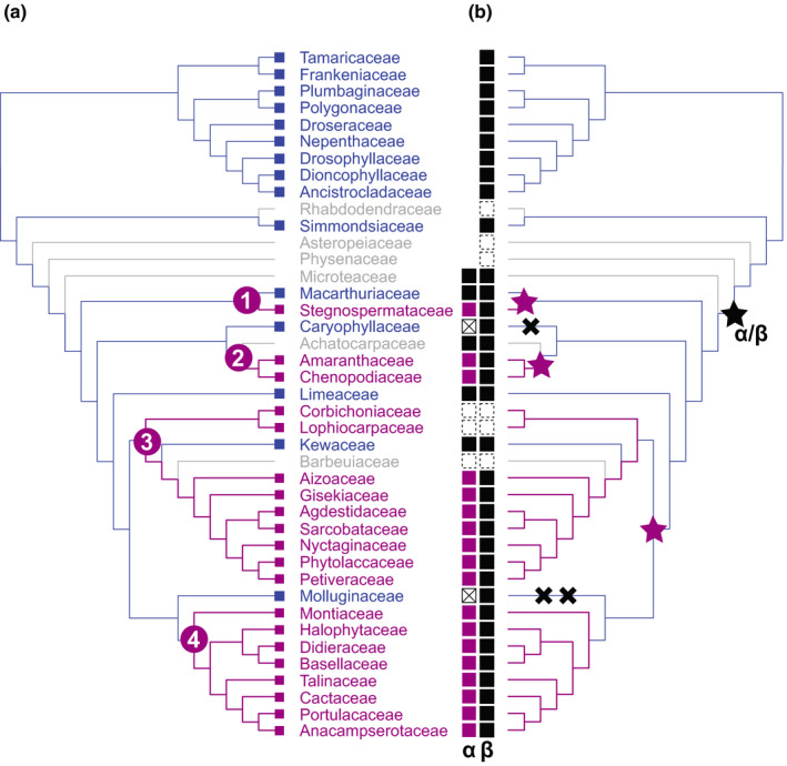
Figure 8.

Summary of the major evolutionary changes inferred with respect to transitions in pigment type and corresponding transitions in l‐DOPA 4,5‐dioxygenase activity. (a) Simplified pigment reconstruction shows family‐level relationships with numbers in purple circles representing the four inferred origins of betalain pigmentation. Tips are coloured according to pigmentation state: anthocyanin (blue), betalain (pink) and unknown (grey). (b) Mirror image of pigment reconstruction topology. A black star marks the initial DODA α/DODA β duplication. Purple stars indicate inferred phylogenetic locations of DODA α gene duplications giving rise to paralogues with high l‐DOPA 4,5‐dioxygenase activity. Black crosses indicate loss of a DODAα paralogue. Tips show inferred presence or absence of DODA α/DODA β: black squares indicate presence of DODA α/DODA β, white squares with a cross indicate loss of DODA α, and white squares with a dashed boundary indicate missing data; DODAα squares coloured purple are inferred to have high L‐DOPA 4,5‐dioxygenase activity.
