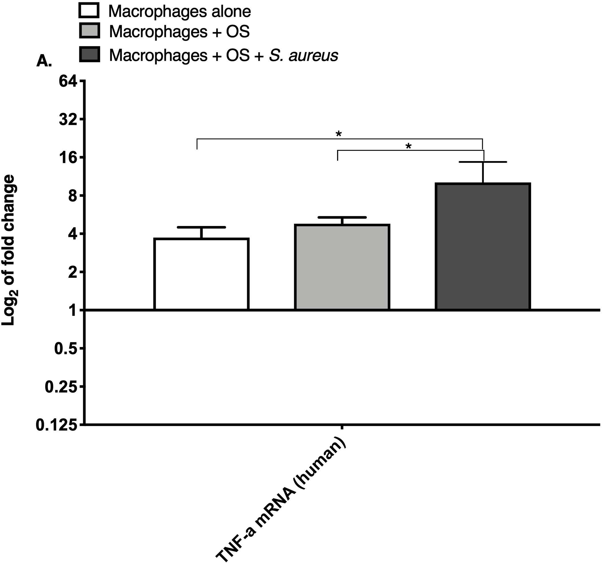
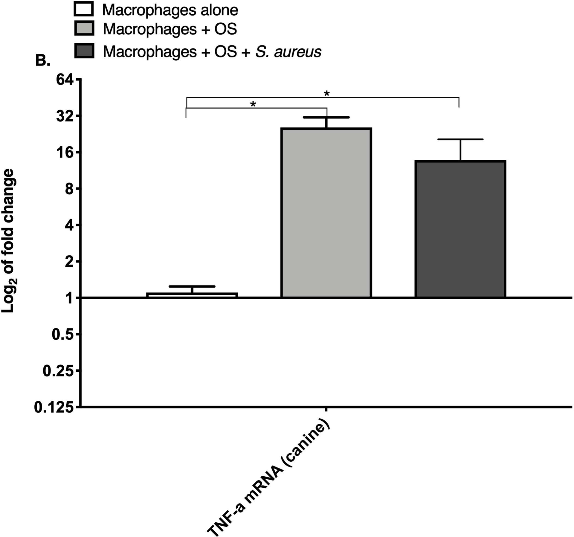
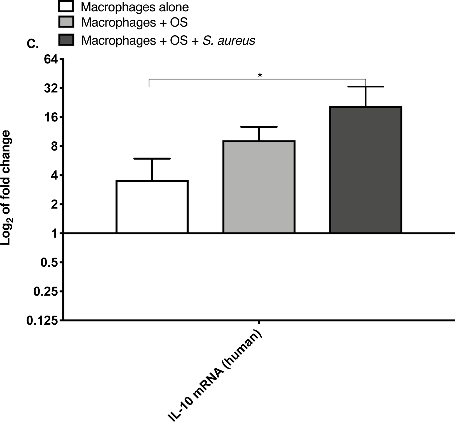
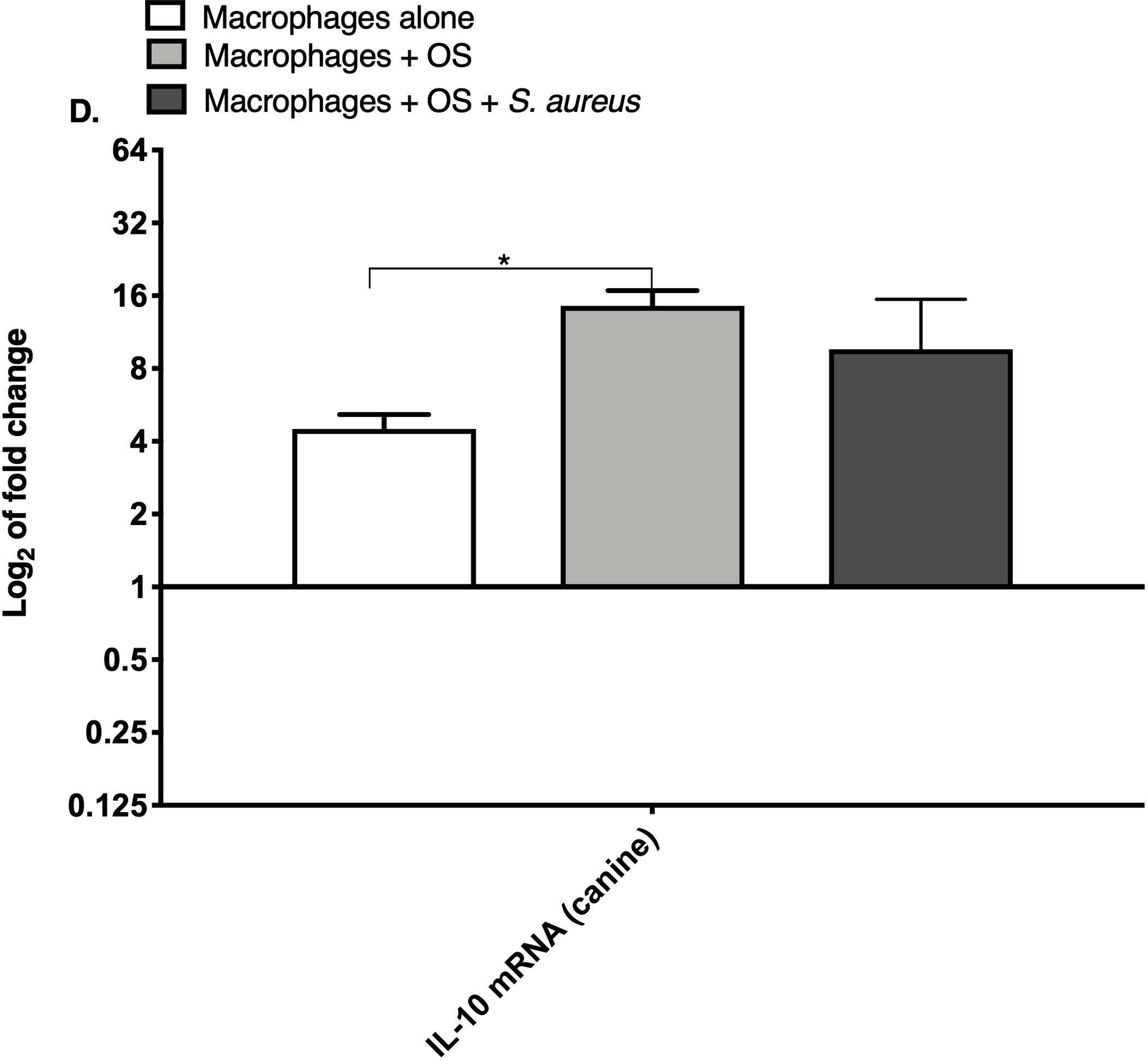
Figure 1: Human and canine macrophage increase inflammatory mRNA profile in the presence of OS and S. aureus.


Bar graphs displaying the level of mRNA expression of (A) TNF-α in human macrophages cultured alone (n = 5), macrophages co-cultured with OS (n = 8), and macrophages co-cultured with OS + S. aureus (n = 9); (B) TNF-α in canine macrophages cultured alone (n = 12), macrophages co-cultured with OS (n = 14), and macrophages co-cultured with OS + S. aureus (n = 10); (C) IL-10 in human macrophages under the same culture conditions as (A); and (D) IL-10 in canine macrophages under the same culture conditions as (B). TNF-α mRNA expression was increased in macrophages co-cultured with OS and S. aureus compared to macrophages cultured alone (p = 0.0105) and to macrophages co-cultured with OS (p = 0.0368). TNF-α mRNA expression was significantly increased in canine macrophages co-cultured with OS (p<0.0001), and in macrophages co-cultured with OS and S. aureus (p = 0.0057) compared to macrophages cultured alone. IL-10 expression was significantly increased in human macrophages co-cultured with OS and S. aureus compared to macrophages cultured alone (p=0.0025). IL-10 expression was significantly increased in canine macrophages co-cultured with OS compared to macrophages cultured alone (p = 0.0023). Data shown as mean ± SEM of triplicate wells, asterisks indicate statistical significance (p<0.05).
