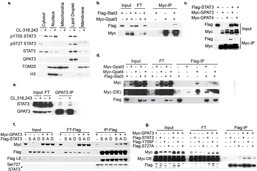
Extended Data Fig. 7: STAT3/GPAT3 interaction.
a. Western blot analysis of fractionated 3T3-L1 adipocytes treated with 10 μM CL-316,243 or vehicle control for 60 min. b.-d. and f. Western blot analysis of input, flow through and immunoprecipitation using Myc-antibody coated beads (b, c) or Flag-antibody coated beads (d, f) of HEK293T cell lysates overexpressing Flag-tagged STAT3 and/or Myc-tagged GPAT3/GPAT4. Blots are representative of three independent replicates. Dark exposure (D.E.). e. Western blot analysis of input and immunoprecipitation using GPAT3 antibody in 3T3-L1 differentiated adipocytes treated with 10 μM CL-316,243 or vehicle control for 15 min. f. Western blot analysis of input, flow through and immunoprecipitation using Flag antibody coated beads of HEK293T cell lysates overexpressing Flag-tagged STAT3 (WT/S727A/S727D) and/or Myc-tagged GPAT3. Blots are representative of three independent replicates. Arrow indicates expected size of Ser727 phosphorylated STAT3; the band observed in the IP samples is a larger non-specific band. g. Western blot analysis of input, flow through, and immunoprecipitation using Flag antibody coated beads from 3T3-L1 differentiated adipocytes with lentiviral overexpression of flag-tagged STAT3 (WT/Y705F/S727A) and/or Myc-tagged GPAT3, cells treated with 10 μM CL-316,243 or vehicle control for 60 min before harvest and IP. These experiments were repeated independently twice with similar results.

