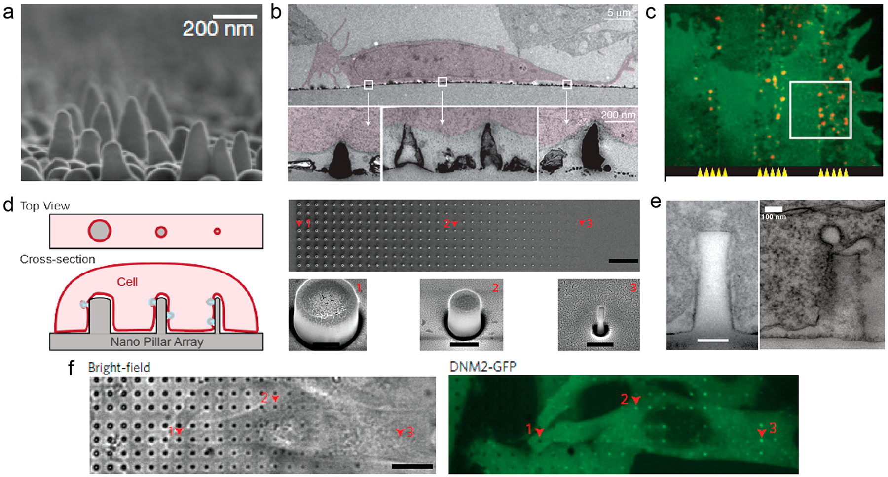
Figure 3.

Vertical nanostructures induce local accumulation of proteins. (a) SEM image of the nanocone substrate.34 (b) TEM images of 3T3 cells on nanocones.34 (c) Fluorescent images show nadrin-2 (red) preferentially accumulated on nanocone strips compared with flat strips.34 (d) (left) Schematic illustration of gradient nanopillars deforming the plasma membrane. (right) SEM images of the gradient nanopillars.29 Scale bars, 10 μm (top), 400 nm (bottom). (e) TEM images of the membrane–nanopillar interface (left) and clathrin-coated pits (right) on the nanopillars.29 Scale bar, 100 nm. (f) A time-averaged image of dynamin2–GFP demonstrates that dynamin–GFP exhibits strong preference to sharp nanopillars.29 Scale bar, 10 μm. Reprinted with permission from the following: ref 34, Copyright 2012 Nature Publishing Group; ref 29, Copyright 2017 Nature Publishing Group.
