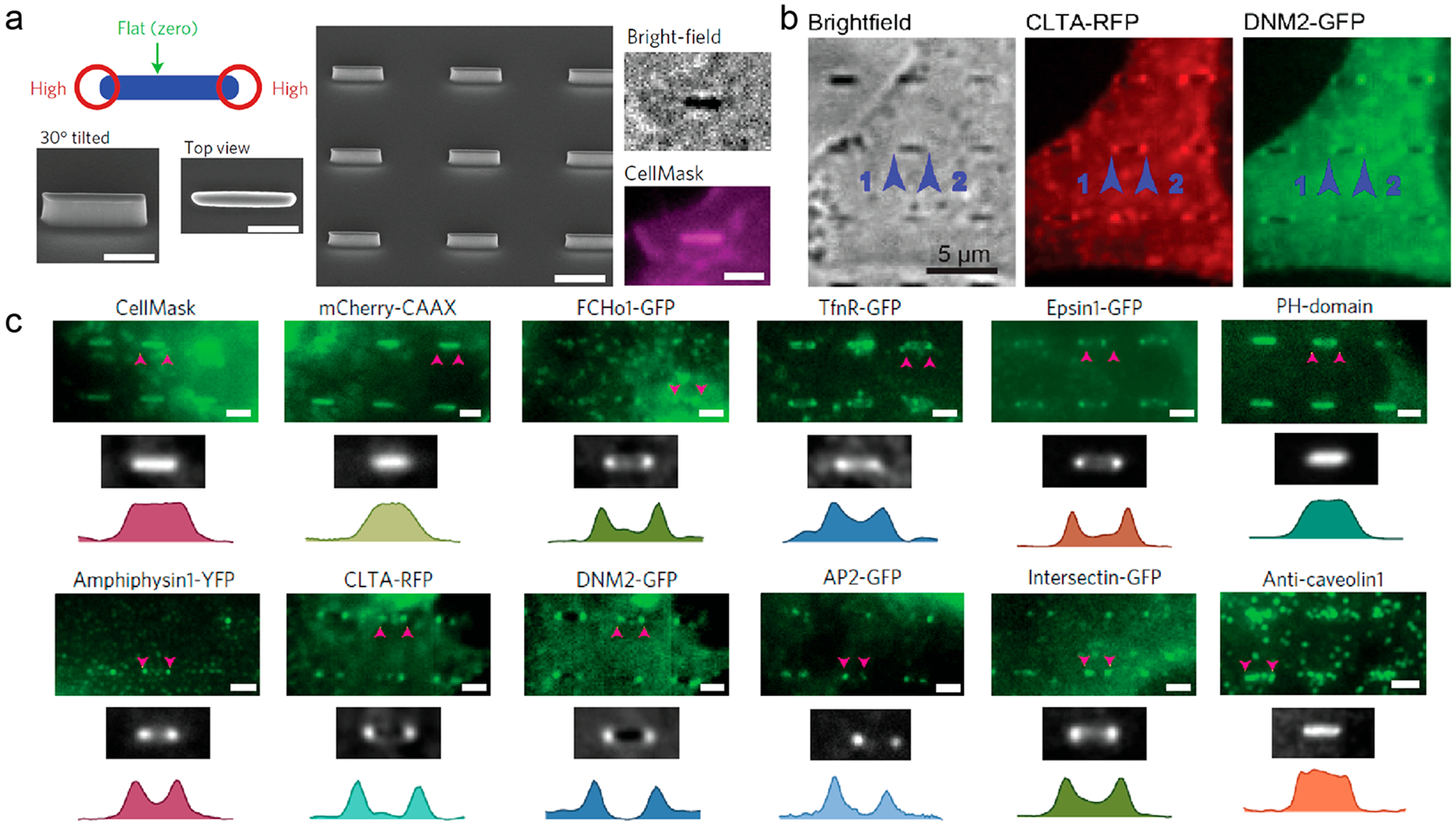
Figure 4.

Clathrin-dependent endocytic proteins accumulate at nanobar ends in a curvature-dependent manner.29 (a) (left and middle) Schematic and SEM images of nanobar structure. Scale bars, 1 μm. (right) CellMask Deep Red staining of SK-MEL-2 cells on the nanobar arrays. Scale bar, 2 μm. (b) Averaged fluorescence images show that clathrin and dynamin-2 preferentially accumulate around the ends of nanobar structures. Scale bar, 5 μm. (c) The distribution of CellMask, mCherry-CAAX, and various endocytic proteins on the nanobar arrays. Scale bars, 2 μm. Reprinted with permission from ref 29. Copyright 2017 Nature Publishing Group.
