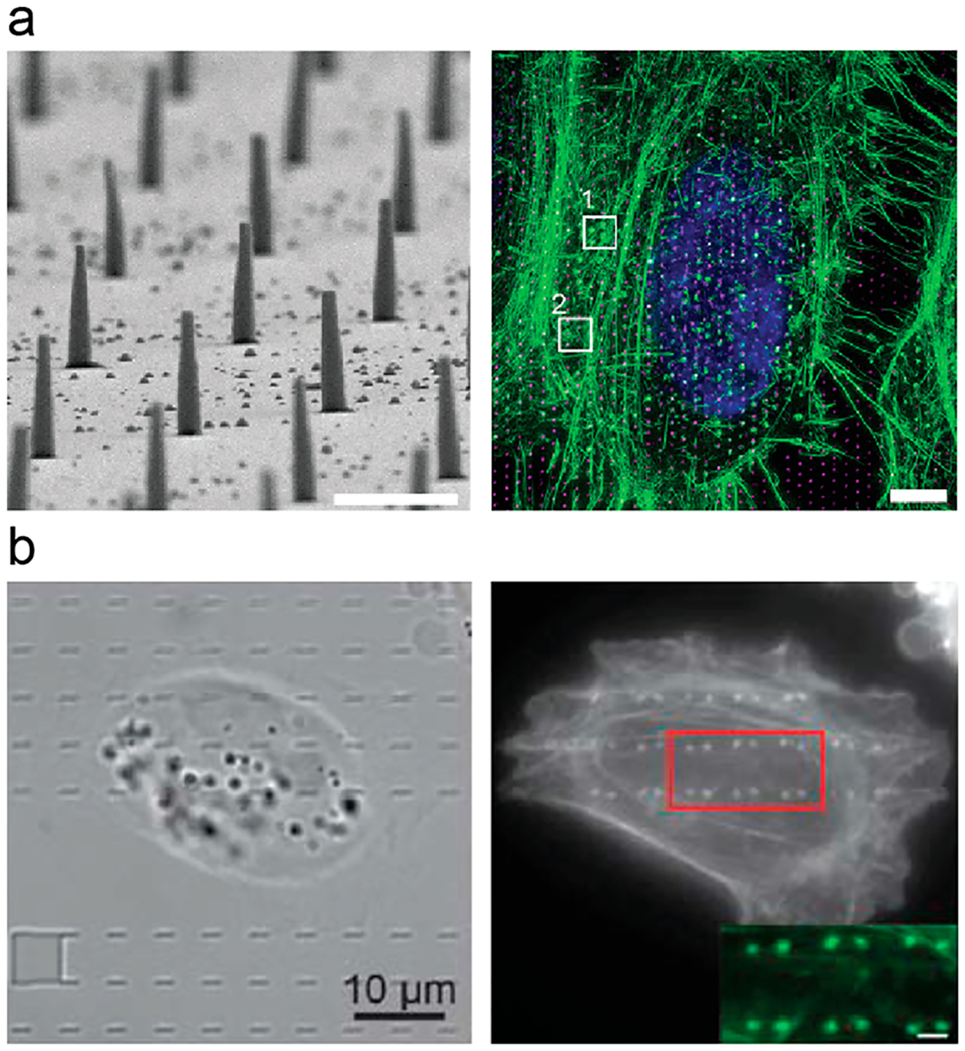
Figure 5.

Vertical nanostructures induce local polymerization of F-actin. (a) (left) SEM image of SU-8 nanopillar arrays. Scale bar, 1 μm. (right) Maximum intensity projection of a 3D-SIM stack showing phalloidin–Alexa488-labeled F-actin (green), Hoechst 34580 labeled nucleus (blue), and 1 μm spaced nanopillars (magenta) in a hexagonal array.37 Scale bar, 5 μm. (b) Full frame images of F-actin shows its strong preference on two ends of nanobars, suggesting a curvature effect.29 Reprinted with permission from the following: ref 37, Copyright 2015 The Royal Society of Chemistry; ref 29, Copyright 2017 Nature Publishing Group.
