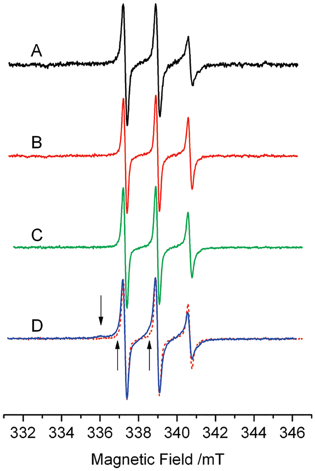
Figure 3.

EPR spectra of 20 μM 9 at 277 K. (A) 9 in buffer. (B) 9: DNA at molar ratio 1:5. (C) 9:Top1 at molar ratio 1:1. (D) 9:DNA: Top1 at molar ratio 1:5:1; this spectrum shows an additional spectral component, absent in spectra A–C, highlighted by the arrows and by the superposition of spectrum B in dashed line.
