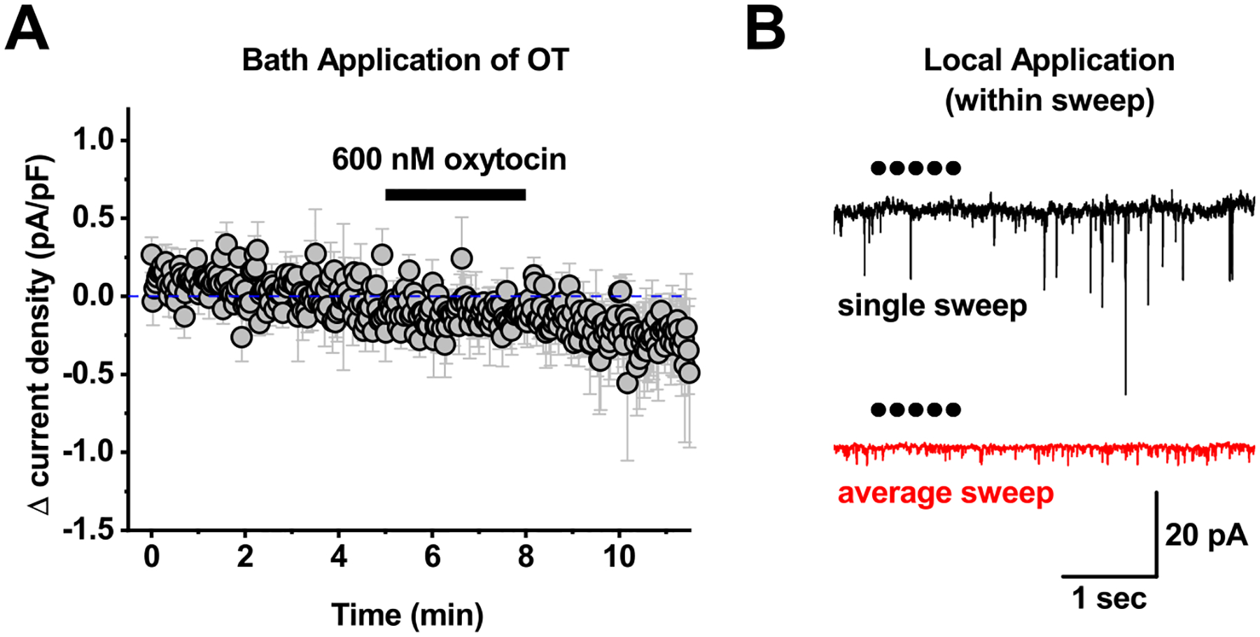
Figure 7: Acute bath and local application of oxytocin to PVN CRH neurons.

A) Bath application of 600 nM oxytocin failed to produce any clear response in PVN CRH neurons voltage clamped at −50 mV. B) For local application experiments, 1 μM oxytocin was loaded into a glass pipette, which was connected to a picospritzer. The tip of the pipette was then placed within ~20 um of the cell soma, and 5–10 msec puffs of oxytocin were delivered at 5 Hz every 30 seconds. The top trace in panel B illustrates a single sweep recorded during one such application. Each dot represents a single 10 msec puff. The bottom trace is an average of 12 sweeps delivered at 30 second intervals from the same cell as illustrated in the top trace. This experiment was repeated 6 times, and none of the 6 PVN CRH neurons tested showed any rapid response to local application of OT. Specifically, when data in each sweep was baseline subtracted to a 1.9 second period immediately before local application, mean current observed in the 6 seconds immediately after local application was identical to baseline (0.08 ± 1.19 pA, n-6, p=0.7).
