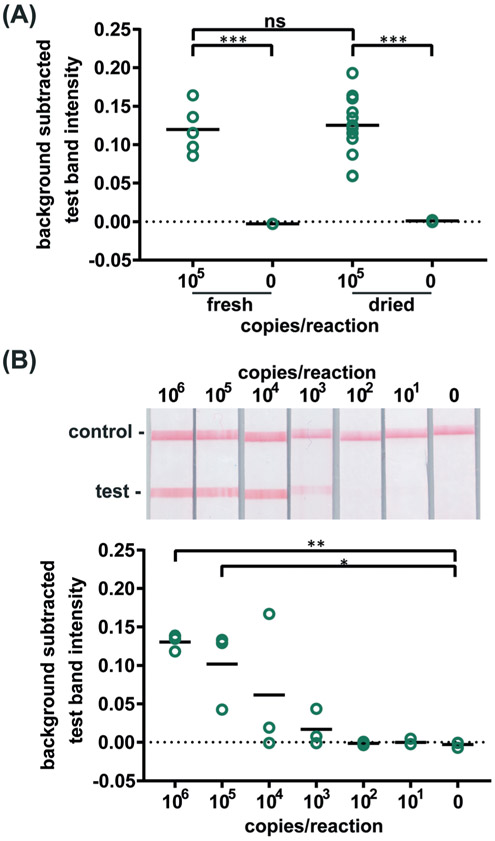
Figure 3. Detection of HIV virus amplified by dried RT-LAMP reagents.
(A) There is no significant difference in test line intensity of labeled amplification products detected on LFIAs after amplification with fresh RT-LAMP reagents as with reagents dried for 21 days. n=5 (fresh), n=13 (dried), circles indicate replicates; *** indicates p ≤ 0.001 (B) Labeled RT-LAMP amplification products are visually detectable from as few as 1,000 HIV virus particles when reactions contain 16% serum. Electrophoresis gels verifying amplification (top, contrast increased for visualization), LFIA test results (middle), and LFIA test line quantification (bottom). n=3, circles indicate replicates; ** indicates p ≤ 0.01; * indicates p ≤ 0.05.

