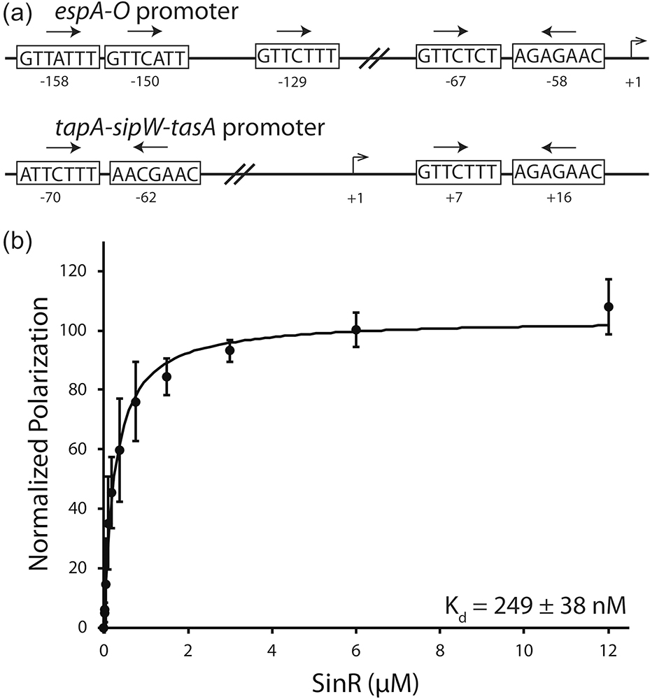
Fig. 6.
Examination of the SinR-DNA interaction. (a) Arrangement of the SinR binding motifs in epsA-O and tapA-sipW-tasA promoters of B. subtilis. SinR binding sequences are shown in boxes. Arrows denote the orientation of the binding sequence. The number of base pairs between binding sequences is noted between the boxes. Transcription start site is labeled at +1 with relative locations of binding sites labeled below each box. (b) Fluorescent anisotropy analysis of SinR binding to a 6-FAM 5ʹ-labelled DNA substrate based on the +7 and +16 tapA-sipW-tasA promoter sequences.

