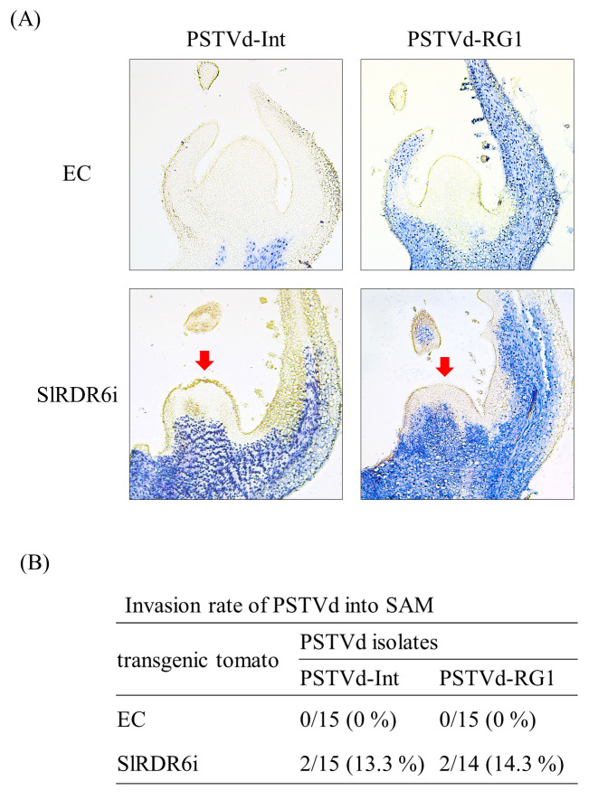
Fig 3. SlRDR6-suppression allowed PSTVd to invade basal part but not apical part of SAM.
(A) Presence of PSTVd in shoot apices of transgenic ‘Moneymaker’ tomato plants was analyzed by in situ hybridization with DIG-labeled cRNA probe for PSTVd at 30–35 dpi. The blue-violet signal in the longitudinal section of shoot apices indicates the presence of PSTVd. PSTVd-Int and PSTVd-RG1 were detected in the basal part but not apical part of SAM in SlRDR6i plants (indicated with a red arrow). (B) Rates of PSTVd invasion into the SAM of EC and SlRDR6i plants. Shoot apices (14 or 15) in each test section were used in the detection of PSTVd. The invasion rates of PSTVd-Int and PSTVd-RG1 into the SAM of SlRDR6i plants were similar.

