Figure 1.

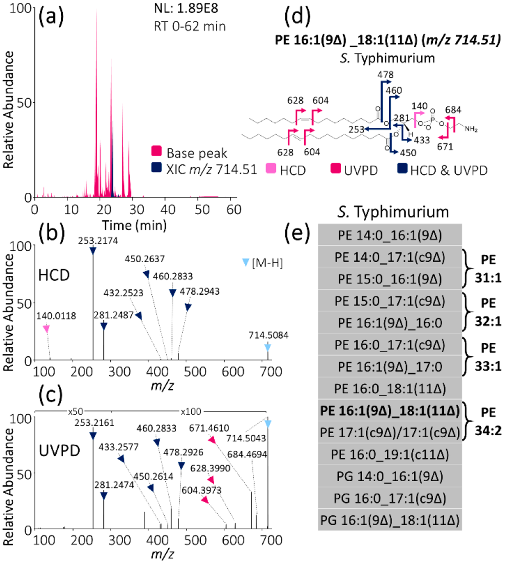
MS analysis of unsaturated GPLs from bacterial extracts: (a) Base peak LC-MS trace of Salmonella enterica serovar Typhimurium (S. Typhimurium) lipid extract with XIC of m/z 714.51 highlighted. (b) HCD mass spectrum of m/z 714.51. (c) 193 nm UVPD mass spectrum (eight pulses, 2.5 mJ per pulse) of m/z 714.51. (d) Fragment ion map of m/z 714.51 identified as PE 16:1(9Δ)_18:1(11Δ). (Fragmentation sites are color-coded to correspond to the ions identified in panels b and c.) (e) List of all identified unsaturated GPLs in S. typhimurium lipid extract using LC-MS/MS with alternating HCD and UVPD.
