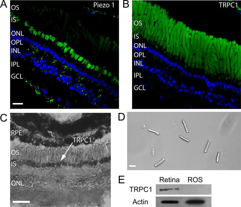
Fig 7. Expression of mechanosensitive channels in the X. laevis retina.
(A) Immunofluorescence for Piezo1 in green and DAPI in blue. (Scale bar is 20 μm for images A, B, and D.) (B) Immunofluorescence for TRPC1 in green and DAPI in blue. (C) Immunohistochemistry for TRPC1. (Scale bar, 50 μm.) (D) Isolated OS obtained by sucrose centrifugation. (E) WB for TRPC1 from the whole retina and isolated ROS as those shown in D. GCL, Ganglion Cell Layer; INL, Inner Nuclear Layer; IPL, Inner Plexiform Layer; IS, inner segment; ONL, Outer Nuclear Layer; OPL, Outer Plexiform Layer; OS, outer segment; ROS, Rod outer segment; RPE, Retinal Pigment Epithelium; TRPC, transient receptor potential canonical; WB, western blot.

