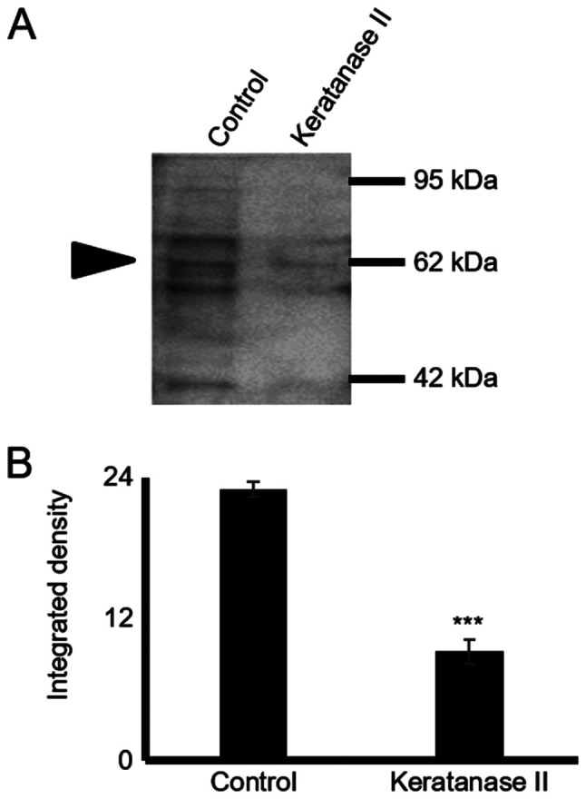
Figure 2.

Glycosylation pattern of lumican secreted by HTB94 cells. (A) Following protein extraction from the culture medium, equal amounts of protein were treated with keratanase II for 16 h at 37°C and blotted with the antibody against keratan sulfate (cloneBD4). Staining with an anti-KS antibody is specific for the KS chains. (B) Protein bands were densitometrically measured utilizing the ImageJ program and expressed as arbitrary units. The position of the nearest respective protein marker band is depicted to the right. Results represent the average of 3 separate experiments. Representative plots are presented. Data are the means ± SEM; ***P≤0.001, statistically significant difference compared with the respective control samples. KS, keratan sulfate.
