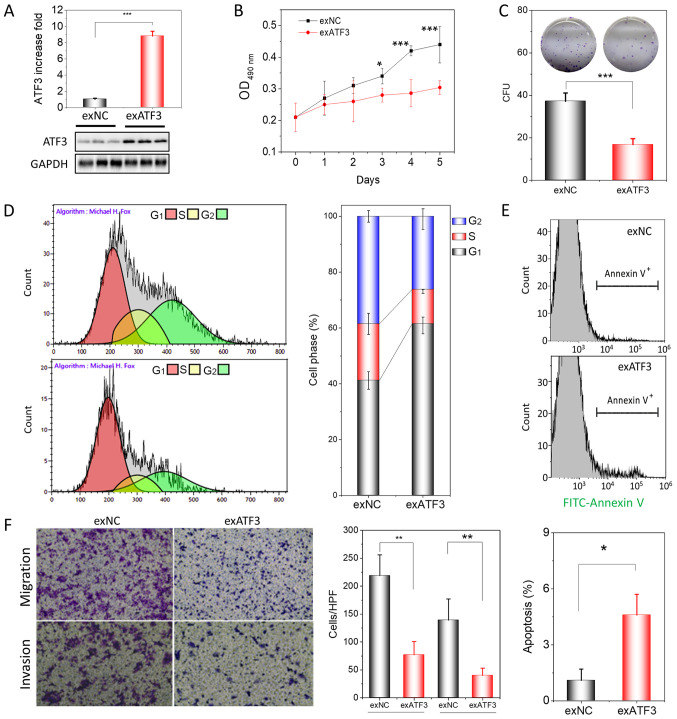
Figure 2.
ATF3 overexpression inhibits proliferation, migration and invasion of HEC-1B cells. (A) Assessment of ATF3 protein expression in exATF3 cells after stable transfection. (B) Assessment of the cell proliferation of exNC and exATF3 cells. (C) Colony-formation assays of exATF3 and exNC cells, and graphical representation of the numbers of colonies in three independent experiments. (D) Cell cycle determined using flow cytometry and the quantitative analysis. (E) Representative images of flow cytometry analysis of apoptotic rates of exNC and exATF3 cells. The apoptotic cells were stained with FITC-Annexin V. (F) Cell migration and invasion assay of exNC and exATF3 cells, and graphical representation of the numbers of migrated and invaded cells in three independent experiments (magnification, ×40). Data are presented as the mean ± SD (n=3) and were analyzed using one-tailed Student's t-test. *P<0.05, **P<0.01, ***P<0.001 vs. controls. ATF3, activating transcriptional factor 3; exATF3, exogeneous ATF3 high expression cells; OD, optical density; NC, negative control; exNC, negative control cells of exATF3; HPF, high-power field; CFU, colony forming unit.

