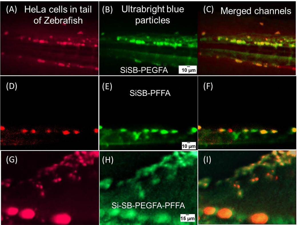
Figure 3.

Co-localization of tumors and folate-functionalized UBMS nanoparticles. Zebrafish injected with red fluorescent HeLa cells in the yolk (A, D and G). Ultrabright blue fluorescent particles functionalized with PEGFA-PEG (B), PFFA (E) and PEG-FA-PFFA (H) injected close to the eye of zebrafish. Corresponding co-localization images of red fluorescent cancerous cells and particles injected in zebrafish (C, F and I). Brightness and contrast of the particles images was optimized for better viewing while keeping same values for all images. The images were taken after ~40 minutes past the particle injection.
