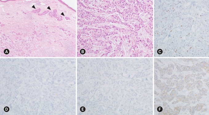
Fig. 2.
Representative histological features and immunohistochemical (IHC) findings of the metastatic carcinoma. The metastatic tumor shows the histologic grade 3, according to the modified Nottingham grading system (tubule formation 3, nuclear pleomorphism 3, and mitotic activities 2), and frequent lymphovascular emboli (A, arrowheads) within the dermal area (A, B). The tumor cells shows 10% Ki-67 labeling index (C), loss of estrogen receptor (D), and progesterone receptor (E). The expression of HER2 was classified as 2+ (F) on IHC, subsequently proven HER2 negativity on HER2 silver in situ hybridization (not shown) (hematoxylin and eosin stain, ×40 [A, B]; IHC stain, ×200 [C−F]).

