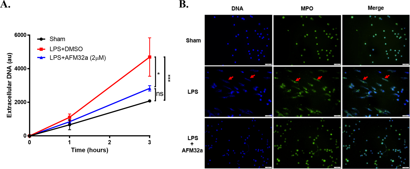
Figure 4. PAD2 inhibition decreased NET formation in vitro.

(A). The graph showed the levels of extracellular DNA in supernatant of cultured mouse neutrophils 1 h and 3 h after LPS treatment (100 ng/ml) with or without AFM32a (2 μM). Extracellular DNA were significantly reduced by AFM32a compared to Group LPS + DMSO. (B). Represent results of immunofluorescence staining showed that AFM32a significantly prevented NET formation. DNA was stained with DAPI (blue), MPO were stained with rabbit anti-mouse MPO antibody and Alexa Fluor 488-conjugated goat anti-rabbit IgG antibody (Green). NETs were recognized as scattered DNA co-localized with MPO (red arrows). Scale bar: 50 μm. DMSO: dimethyl sulfoxide; LPS: lipopolysaccharide; MPO: myeloperoxidase; au: arbitrary units; ns: no significance; * P <0.05; *** P <0.001.
