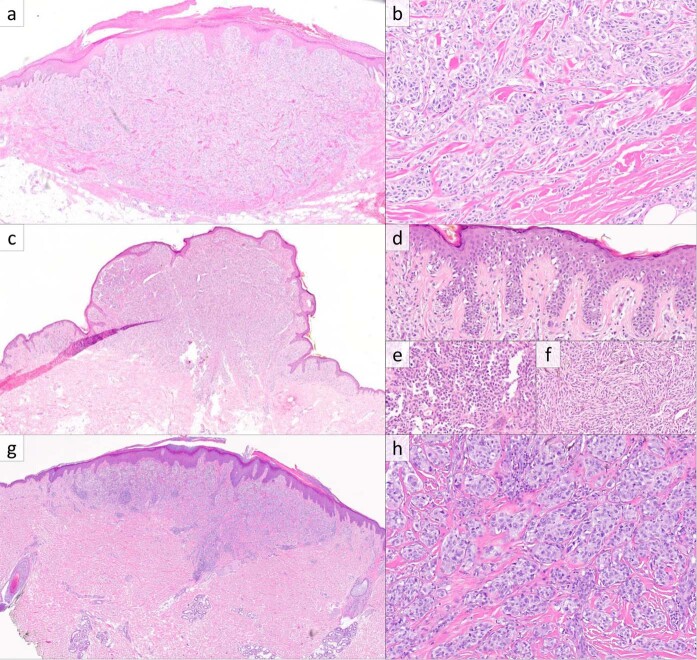
Fig. 1. RAF1-fusion melanoma, primary cutaneous lesions.
a Histopathologic examination of case 2 reveals a melanocytic neoplasm with a slightly domed surface and wedge-shaped base centered in the dermis (H&E, ×20). b The deep aspect of case 2 consists of nested large epithelioid melanocytes with associated dense collagen fibers and scattered mitotic figures (H&E, ×200). c Case 3 showed an exophytic component with a plaque-like growth pattern at the periphery (H&E, ×20). For case 3, the radial growth phase at the periphery showed intraepidermal growth with pagetoid scatter (d), while the vertical growth phase contained predominantly epithelioid melanocytes (e) with a deep zone of fascicular growth of spindled cells with focal cytoplasmic pigmentation (f) (H&E, ×200, ×400, and ×400). g, h Case 8 showed a melanocytic neoplasm with a domed surface and wedge-shaped base with nested growth in the deep aspect with dense collagen fibers and scattered mitoses (H&E, ×20 and ×200).

