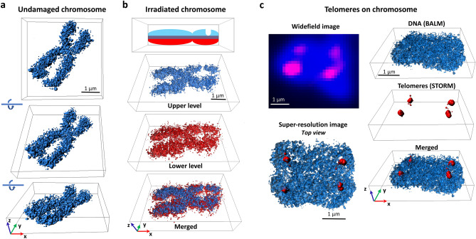
Figure 3.
3D super-resolution imaging of metaphase chromosomes. (a) Three-dimensional BALM images of an undamaged human metaphase chromosome. (b) 3D BALM images of an irradiated chromosome. A schematic representation of irradiated chromosome with a groove on one of the arms and three-dimensional surface plots at the upper (blue), and lower (red), layers. (c) (Top left) Wide-field image of a SxO-stained human chromosome (blue) and telomeric regions on the chromosome (magenta). (Bottom left) A top view of merged re-constituted super resolution (BALM) image of the chromosome and re-constituted super resolution (STORM) image of the telomeric regions of this chromosome. (Right) Three-dimensional surface plots of BALM (blue), STORM (red), and both images superimposed.

