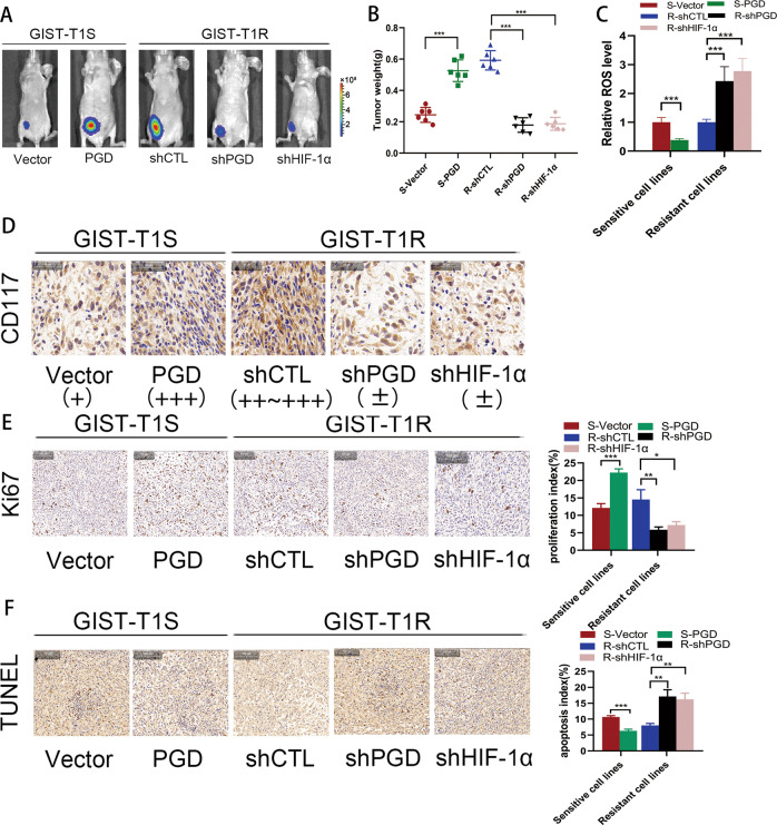
Fig. 6. HIF-1α promotes drug resistance in vivo.
a Bioluminescent images of mice administered with GIST cells. Images were taken after 8 weeks. n = 3 per group. b The in vivo effect of HIF-1α and PGD was evaluated in xenograft mouse models bearing tumors originating from GIST-T1 and GIST-882 cells; n = 3 per group. c ROS levels were measured in xenograft mouse models bearing tumors originating from GIST-T1 and GIST-882 cells; n = 3 per group. d Representative CD117 staining of primary tumor tissues. e Representative Ki67 staining of primary tumor tissues. f TUNEL assay were used to determine the effects of HIF-1α and PGD expression alteration on cell apoptosis in the samples collected from nude mice. The error bars represent the mean (n = 3) ± S.D. *P < 0.05, **P < 0.01, ***P < 0.001.

