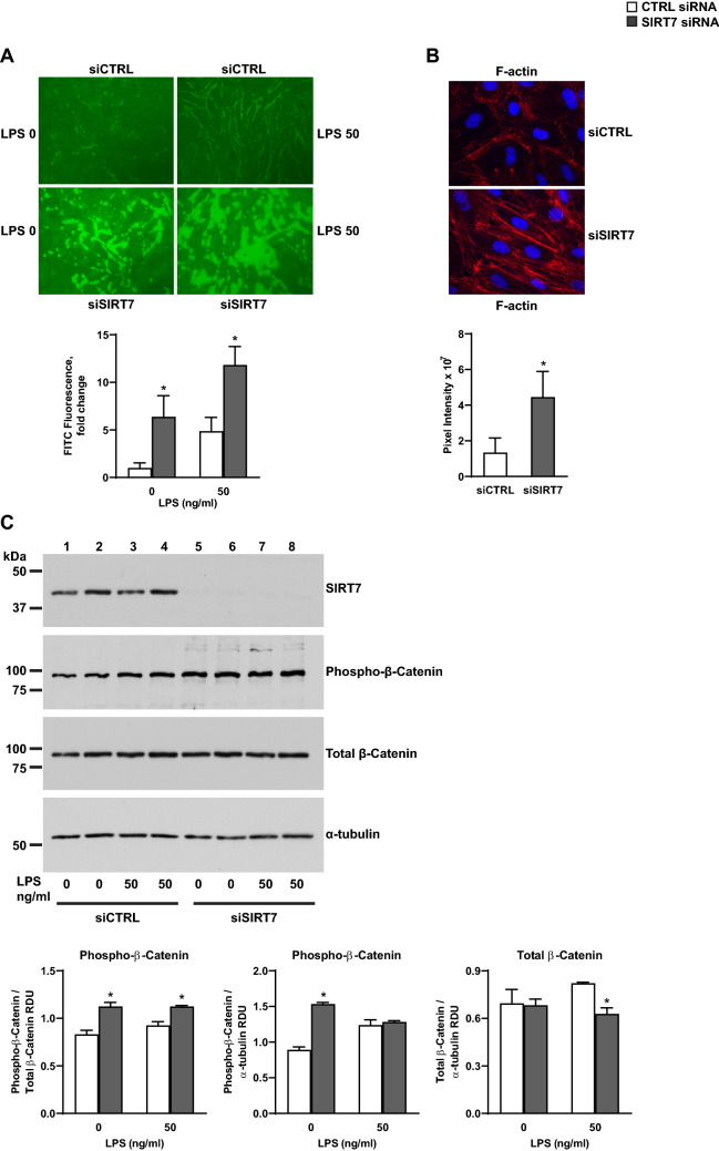
Figure 7.
Effects of SIRT7 silencing on barrier permeability in endothelial cell cultures. (A) Effect of SIRT7 silencing in HPAEC on endothelial barrier permeability measured by XPerT assays 6 h after stimulation with LPS 50 µg/ml or no stimulus. Average fluorescence values and standard deviations of 6 images expressed as fold changes relative to the average value of unstimulated, CTRL-silenced cultures are shown below the image. (B) Representative IF images for F-actin stress fibers in CTRL- or SIRT7-silenced HPAEC 48 h after transfection. Average pixel intensities and standard deviations of 6 images per condition are shown. (C) Phosphorylated and total β-Catenin protein levels 6 h after LPS stimulation or no treatment in CTRL-silenced (□) or SIRT7-silenced (■) HPAEC cultures. Densitometry measurements of phosphorylated β-Catenin relative to total β-Catenin or α-tubulin (also shown in Supplementary Figure 11E) and total β-Catenin relative to α-tubulin (shown in Supplementary Figure 11C) are shown. Each bar represents the average of two separate samples per condition. Significant differences (P < 0.05) are denoted by stars. P values were calculated using the Student’s t test. For (C), different proteins for the same group of samples are demarcated by white spaces.

