Figure 4.
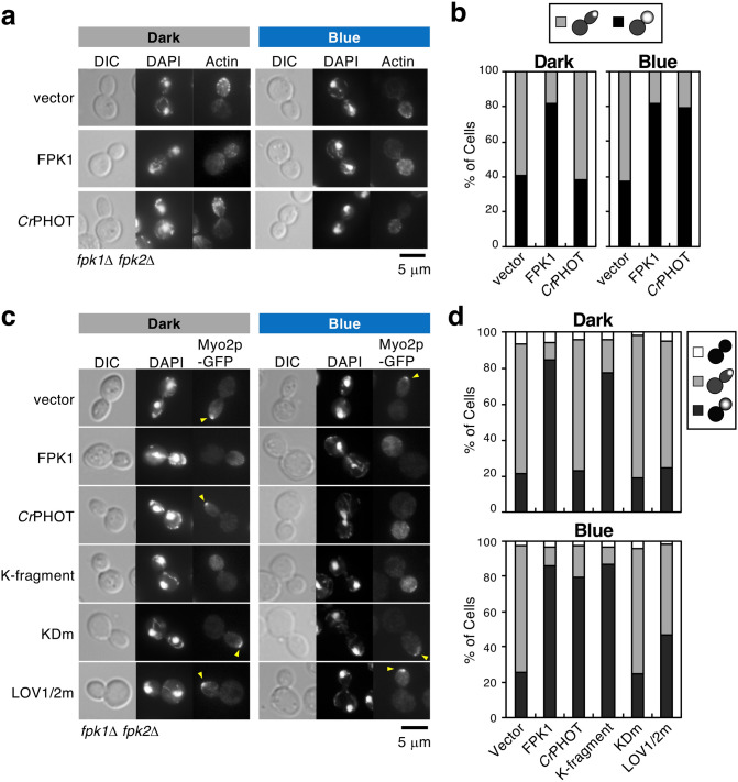
Optical control of actin depolarization associated with apical-isotropic growth switching. (a, b) Optical control of F-actin distribution by CrPHOT. Yeast fpk1Δ fpk2Δ cells carrying pKT1639 (pRS416-FPK1) or pRS416-CrPHOT were cultured in darkness (Dark) or under 10 μmol m-2 s-1 BL irradiation (Blue) at 18 °C, followed by staining with phalloidin-TRITC and DAPI to visualize actin and nuclei, respectively. (a) A representative cell image by microscopic observation. Scale bar = 5 μm. (b) Large-budded cells with divided nuclei were classified as showing actin polarized to the bud tip (grey) or its distribution in whole daughter cells (black). n = 155–253. (c, d) Optical control of Myo2p-GFP localization in a kinase-dependent manner. KKT353 (fpk1Δ fpk2Δ MYO2-GFP) cells carrying pKT1639 (pRS416-FPK1) or pRS416-CrPHOT or its derivatives were cultured in YPDA medium in darkness (Dark) or under BL irradiation (Blue) at 18 °C, followed by staining with DAPI to visualize nuclei. (c) A representative cell image obtained by microscopic observation. Arrowheads indicate Myo2p-GFP polarized to the bud tip. Scale bar = 5 μm. (d) Large-budded cells with divided nuclei were classified as showing Myo2p-GFP distributed in whole daughter cells (black), polarized to the bud tip (grey) or delocalized (white). n = 96–180.

