Figure 9.
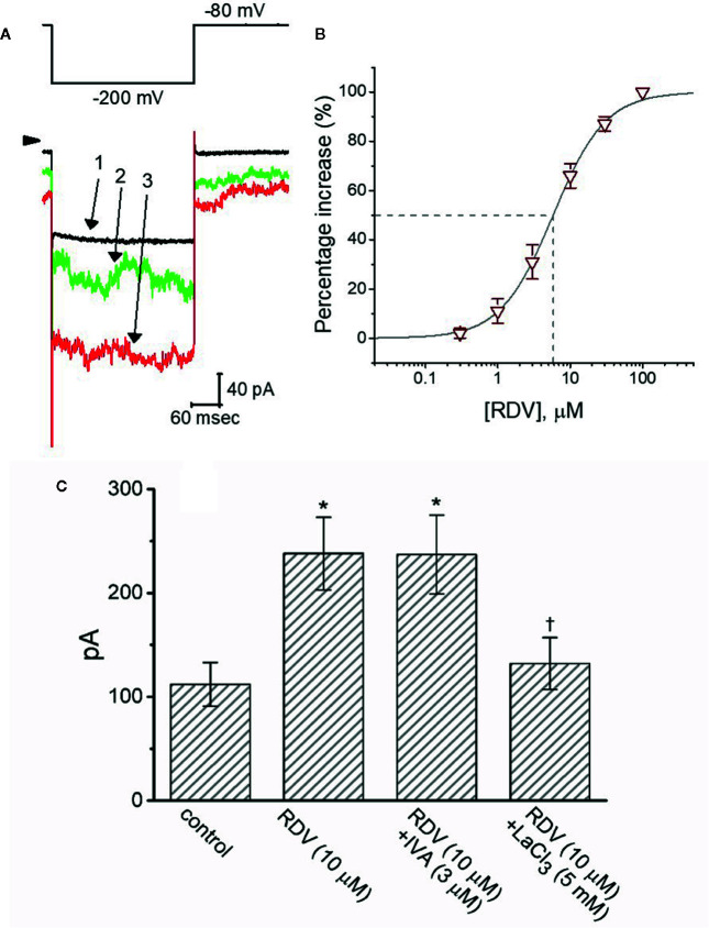
The stimulatory effect of RDV on membrane electroporation-induced current (I MEP) identified in GH3 cells. In this separate set of experiments, we bathed cells in Ca2+-free, Tyrode’s solution and the recording pipette was filled with K+-containing solution. (A) Representative I MEP traces obtained in the control (1) and during cell exposure to 3 μM RDV (2) or 10 μM RDV (3). The voltage-clamp protocol applied is denoted in the upper part, arrowhead is the zero current level and the calibration mark at the right lower part applies all current traces. Noticeably, the addition of RDV causes a measurable increase in the amplitude of I MEP elicited by large membrane hyperpolarization from −80 to −200 mV with a duration of 300 ms. (B) Concentration-dependent stimulation of I MEP produced by RDV in GH3 cells (mean ± SEM; n=8 for each point). Current amplitude was measured at the end of hyperpolarizing pulse from −80 to −200 mV with a duration of 300 ms, and the vertical dashed line is placed at the IC50 value required for RDV-stimulated I MEP. (C) Summary bar graph showing effect of RDV, RDV plus ivabradine (IVA) and RDV plus LaCl3 on I MEP amplitude (mean ± SEM; n=8 for each bar). Current amplitude was taken at the end of hyperpolarizing voltage pulse from −80 to −200 mV with a duration of 300 ms. *Significantly different from control (P<0.05) and Ɨsignificantly different from ZDV (10 M) alone group (P<0.05).

