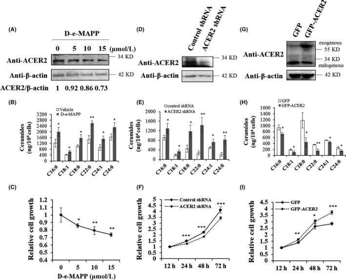
Figure 2.

ACER2 promotes HCC cell proliferation. Huh‐7 cells were treated with D‐e‐MAPP at the indicated concentrations for 24 h (A‐C). Cell lysates were subjected to western blot with indicated antibodies, and the relative protein level of ACER2 was quantified by ImageJ software (A). Intracellular ceramide was detected by UHPLC‐QTOF‐MS (B). Cell proliferation was investigated using the CCK8 assay (C). Lentivirus containing plasmid encoding ACER2 shRNA (D‐F) or full‐length ACER2 (G‐I) was used to infect Huh‐7 cells for 48 h, and then, cells were treated with puromycin until stable cell lines were established. The efficiency of ACER2 knockdown (D) or overexpression (G) was confirmed by western blot with an anti‐ACER2 antibody. Cells lysates were subjected to ceramide analysis (E, H). Huh‐7 cells with stable ACER2 knockdown (F) or overexpression (I) were seeded, and cell survival was measured at 12, 24, 48, and 72 h using the CCK‐8 assay (F, I). Error bars represent the standard deviation (SD) from 3 independent experiments. Statistical significance was analyzed using Student t test (*P < .05; **P < .01; ***P < .001)
