Figure 3.

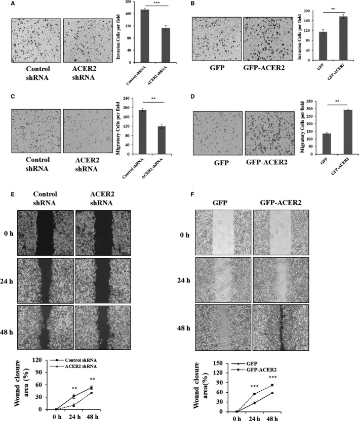
ACER2 promotes HCC cell invasion and migration in vitro. Stable ACER2 knockdown (A, C, E) or overexpressing (B, D, F) cells were seeded. Transwell assays (A‐D) and wound‐healing assay (scale bar: 50 μm) (E, F) were performed in Huh‐7 cells. Cell numbers (A‐D) were counted using ImageJ software. The histograms (A‐D) represent the mean values of invasive and migrated cells per high‐power field (from at least 5 fields, mean ± SD) (** P < .01; *** P < .001). The wound closure (E, F) area was quantified by ImageJ software. Error bar represents the SD from 3 independent experiments, and statistical significance was analyzed using Student t test (** P < .01; *** P < .001)
