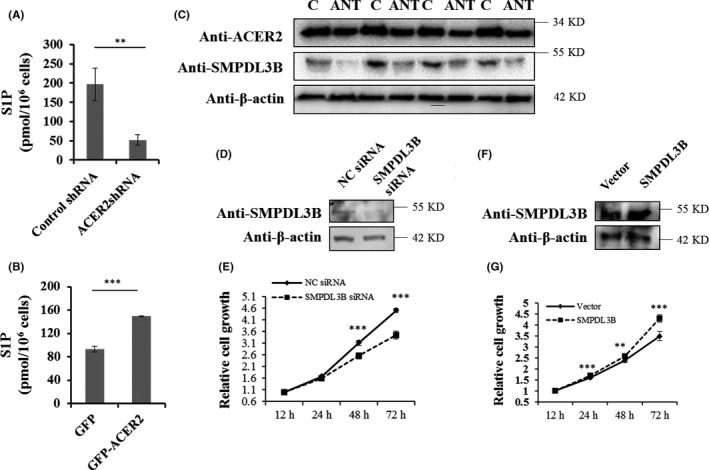
Figure 5.

SMPDL3B promotes HCC cell proliferation. The amounts of S1P in stable ACER2 knockdown (A) or overexpression (B) Huh‐7 cells were measured with an ELISA kit. Error bars represent the SD from 3 independent experiments, and statistical significance were analyzed using Student t test (**P < .01; ***P < .001). C, SMPDL3B expression in HCC human samples, same samples from Figure 1(B) and same images of ACER2 and β‐actin shown in Figure 1(B). Huh‐7 cells with SMPDL3B knockdown (D and E) or overexpression (F and G) were seeded, the protein level of SMPDL3B was measured (D, F) and cell survival at 12, 24, 48, and 72 h was assessed using the CCK‐8 assay (E, G). Error bars represent the standard deviation (SD) from 3 independent experiments. Statistical significance was analyzed using Student t test (**P < .01; ***P < .001)
