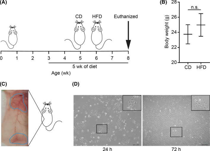
FIGURE 1.

Establishment of mouse mammary epithelial cells (MMECs). A, Schema of feeding of Trp53‐null mice with a high‐fat diet (HFD) or a control diet (CD) for 5 wk. Mice were killed at 8 wk and mammary glands were harvested. B, Body weight of the mice at the time of death (N = 7 for each group). C, Ventral view of abdominal (bottom) and thoracic (upper) mammary glands incised from either side of the mouse. D, MMECs cultured in serum‐free EpiCult‐B medium supplemented with recombinant human (rh) basic fibroblast growth factor, rh epidermal growth factor, heparin, and insulin. Images captured 24 h (left) and 72 h (right) after seeding. Scale bar = 200 µm. Data are mean ± SE. *P < .05 (Student’s t test) n.s., non‐significant
