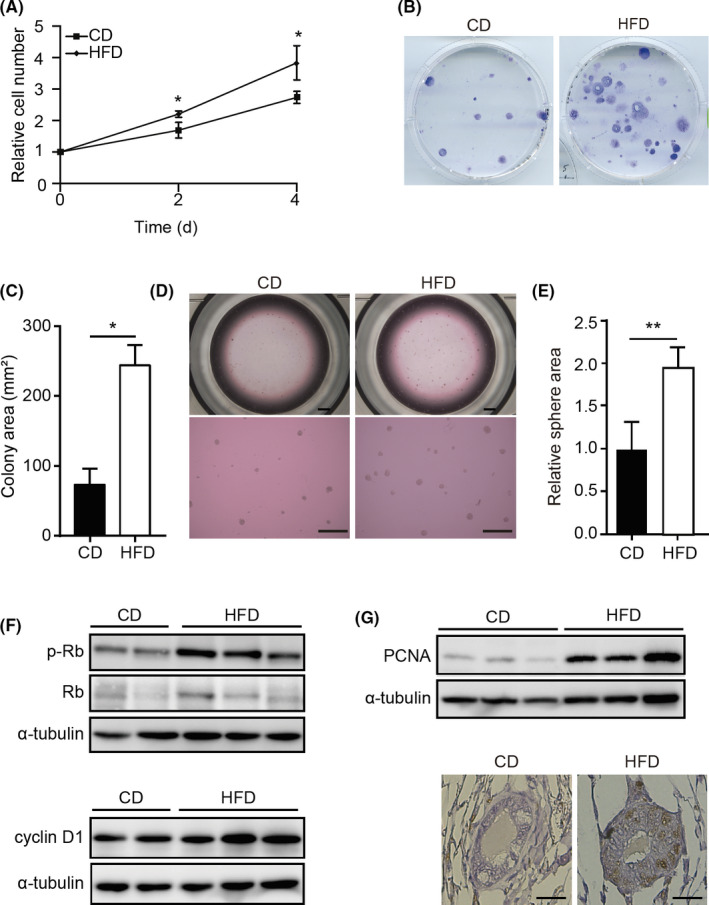
FIGURE 2.

High‐fat diet (HFD) stimulates growth of mouse mammary epithelial cells (MMECs). A, WST‐1‐based cell proliferation assay of MMECs derived from mice fed control diet (CD‐MMECs; N = 3) or HFD (HFD‐MMECs; N = 3) B, Colony formation assay of MMECs derived from mice fed with CD or HFD. C, Quantitative analysis of the colony area (N = 3 for each group). D, Mammosphere formation assay of CD‐MMECs and HFD‐MMECs. Scale bar = 100 µm. E, Quantitative analysis of the sphere area (N = 3 for each group). F, Immunoblotting (IB) of indicated proteins in HFD‐MMECs and CD‐MMECs. Experiments were done in triplicate or duplicate. α‐Tubulin was used as a loading control. G, IB of proliferating cell nuclear antigen (PCNA) in CD‐MMECs and HFD‐MMECs (upper), a representative immunohistochemistry staining of PCNA in the mammary gland tissue in mice fed CD or HFD (bottom). Scale bar = 100 µm. Data are mean ± SE. *P < .05, **P < .01 (Student’s t test). p‐, phosphorylated; Rb, retinoblastoma protein
