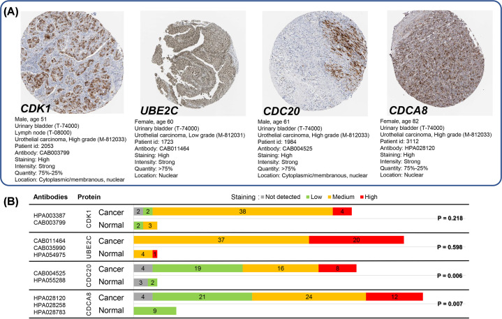
Figure 8. Antibody staining of hub genes in immunohistochemistry images between normal urinary bladder and urothelial cancer tissues of bladder.
(A) High antibody staining immunohistochemistry images of four hub genes are available at https://www.proteinatlas.org/ENSG00000170312-CDK1/pathology/urothelial+cancer# (CDK1), https://www.proteinatlas.org/ENSG00000175063-UBE2C/pathology/urothelial+cancer# (UBE2C), https://www.proteinatlas.org/ENSG00000117399-CDC20/pathology/urothelial+cancer# (CDC20) and https://www.proteinatlas.org/ENSG00000134690-CDCA8/pathology/urothelial+cancer# (CDCA8) in THPA, respectively. (B) Two antibodies were used for CDK1 (HPA003387 and CAB003799) and CDC20 (CAB004525 and HPA055288) and three for UBE2C (CAB011464, CAB035990 and HPA054975) and CDCA8 (HPA028120, HPA028258 and HPA028783). High positive staining rate was detected in urothelial cancer tissues of bladder for CDK1 (44/46), UBE2C (57/57), CDC20 (43/47) and CDCA8 (57/61), however, only CDC20 (P=0.006) and CDCA8 (P=0.007) showed significant results by comparing normal urinary bladder with urothelial cancer tissues of bladder using Mann–Whitney test. The cutoff P-value was set as 0.05.

