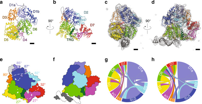
Fig. 3. Cryo-EM reconstruction of dsDNA-bound Mfd reveals a mobile D3 module and the interaction of dsDNA with the motor lobes.
a, b Orthogonal views of nucleotide-free Mfd (PDB ID 2EYQ). Scale bar is equivalent to 10 Å. c, d Orthogonal views of the cryo-EM reconstruction (gray surface) with the fitted Mfd model shown as a cartoon and colored as in a. DNA is shown in gray. e, f Cartoons of Mfd structures in the nucleotide free e and DNA-bound form f after superposition on the Cα trace of D5 and highlighting the relative rotation of Mfd domains in the free and tight DNA-bound state. g, h Chord plots highlighting intramolecular rearrangements in Mfd before g and after h DNA binding. In all panels, Mfd is colored by domain as in Fig. 2a.

