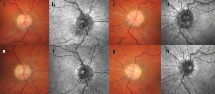
Fig 2.
Progressive optical coherence tomography of retinal structures. This patient presented with papilloedema (a–d) was managed locally and re-referred back to our services following weight loss at 14 months with resolving papilloedema (e–h). The images show the difference between colour fundus images (a, c, e, g) and infrared images (b, d, f, h) taken using Heidelberg Spectralis optical coherence tomography. a) The right optic nerve head showing swelling at presentation. b) Greater fidelity image of the swelling of right optic nerve head. c) The left optic nerve head showing swelling at the presentation. d) Comparative image showing left optic nerve head swelling. e) The right optic nerve head showing resolving of the swelling compared with (a). f) Image showing the reduction in swelling compared with (b). g) The left optic nerve head showing resolving of swelling compared with (c). h) Image showing resolving of swelling compared with (d).

