
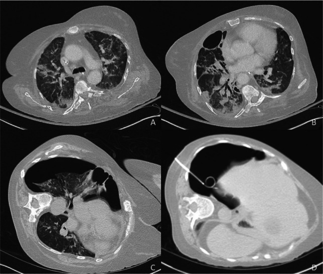
Fig 1.
Computed tomography prior to pneumothorax showing infiltrates in keeping with COVID-19 infection (a, b) and right upper lobe bulla (b), with pneumothorax immediately prior to (c) and immediately after (d) insertion of pleural drain.
Official websites use .gov
A
.gov website belongs to an official
government organization in the United States.
Secure .gov websites use HTTPS
A lock (
) or https:// means you've safely
connected to the .gov website. Share sensitive
information only on official, secure websites.
Computed tomography prior to pneumothorax showing infiltrates in keeping with COVID-19 infection (a, b) and right upper lobe bulla (b), with pneumothorax immediately prior to (c) and immediately after (d) insertion of pleural drain.