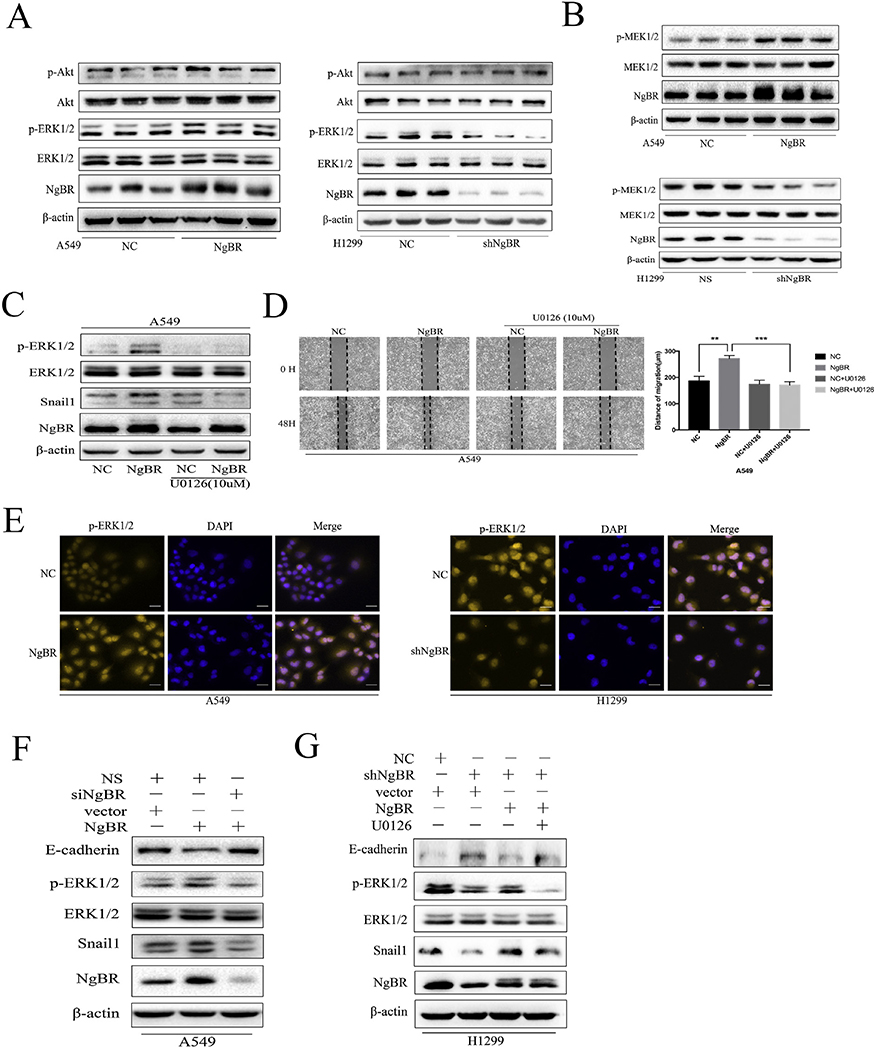
Fig. 5. NgBR expression upregulates Snaill expression through activation of the MEK/ERK/Snaill pathway in NSCLC cells but not the PI3K/Akt pathway.
A, levels of p-ERK1/2 and p-Akt proteins were assessed using Western blotting. Total ERK1/2 and Akt proteins were used as a reference for loading controls in stable A549 and H1299 cells. B, levels of pMEK1/2 protein were assessed using Western blot in stable A549 and HI299 cells. Levels of total MEK1/2 and β-actin proteins were used as loading controls. C, A549 cells were stably transfected with pIRES-NC (NC) and pIRES-NgBR (NgBR) and then treated with or without U0126 (10 μM) for 24 h and then subjected to Western blot analysis of p-ERKl/2, ERK1/2, and Snaill. D, A549 cells were stably transfected with pIRES-NC (NC) and pIRES-NgBR (NgBR) and then incubated with or without U0126 (10 μM) for 24 h and subjected to the wound-healing assay to assess tumor cell migration. Images were taken at 0 h and 48 h. Scale bar, 200 pm (left). Quantitative data of the wound-healing assay (right). Error bar, SD of three independent experiments. **p < .01 and ***P < .001. E, stable A549 and H1299 cells were immunostained with p-ERKl/2 antibody, while cell nuclei were stained with DAPI staining. Scale bar, 37 μm. F and G, stably NgBR or NC transfected A549 cells were transiently transfected with siNgBR or NS for 48 h, while stably shNgBR or NC transfected H1299 cells were transiently transfected with pIRES-NgBR (NgBR) or pIRES-NC (NC) for 48 h. These cells were then subjected to treatment with or without U0126 (10 μM) for additional 24 h and subjected to Western blot analysis of E-cadherin, p-ERKl/2, Snaill protein levels. Total ERK1/2 and β-actin levels were used as loading controls.

