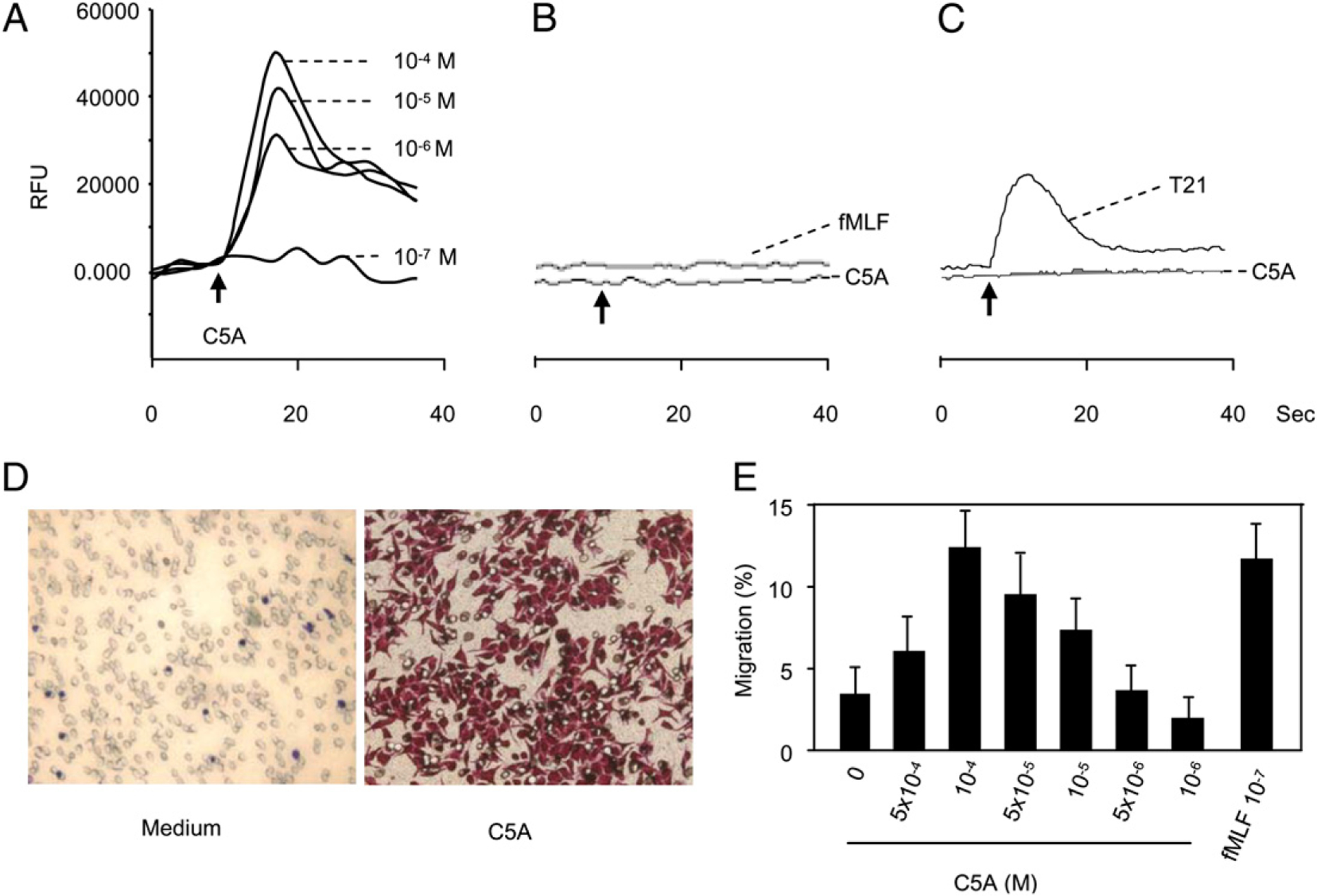
FIGURE 4.

Activation of FPR transfected cells (ETFR cells) by HCV peptide (C5A). Calcium mobilization in ETFR tranfectants (A), rat basophilic leukemia parent cells (B), or FPRL1/293 cells (C) was measured with FlexStation II384 system using Fura-3AM. Changes in intracellular calcium concentration in response to agonists were recorded as relative fluorescence units (RFUs). D and E, Induction of ETFR cell migration by HCV peptide (C5A). Different concentrations of HCV peptide (C5A) were placed in the lower wells of the chemotaxis chamber; cell suspension was placed in the upper wells. The upper and lower wells were separated by polycarbonate filters. After incubation, the cells migrated across the filters were stained and counted or collected and counted by FACSCalibur. D, Migration of ETFR cells across the filters in response to 10−5 M HCV peptide (C5A; original magnification ×200). E, Percentage of ETFR cell migration in response to HCV peptide (C5A) in total loaded cells.
