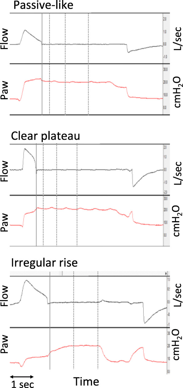
Fig. 2.

Classification of airway pressure waveform pattern during occlusion. Airway flow (in l/s) and airway pressure (Paw, in cmH2O) waveforms representative of the three patterns of Paw during occlusion, from three different patients. The solid vertical line indicates the point of occlusion and subsequent dotted lines the points at 0.3, 1, and 2 s post-occlusion. Each pattern was characterized by the relationships among Paw at different time points relative to the occlusion: occlusion, 0 s (Pawocc), 0.3 s (Paw0.3s), 1 s (Paw1s), and 2 s (Paw2s). Upper panel: a “passive-like” pattern with a rapid decrease in Paw (Pawocc > Paw0.3s), followed by plateau (Paw1s − Paw0.3s < 1 and Paw2s − Paw1s < 1 cmH2O). Middle panel: a “clear plateau” pattern with an early increase in Paw (Pawocc < Paw0.3s), followed by plateau (Paw1s − Paw0.3s < 1 and Paw2s − Paw1s < 1 cmH2O). Lower panel: an “irregular rise” pattern with a slow increase in Paw (Paw1s − Paw0.3s > 1 cmH2O) with plateau (Paw2s − Paw1s < 1 cmH2O)
