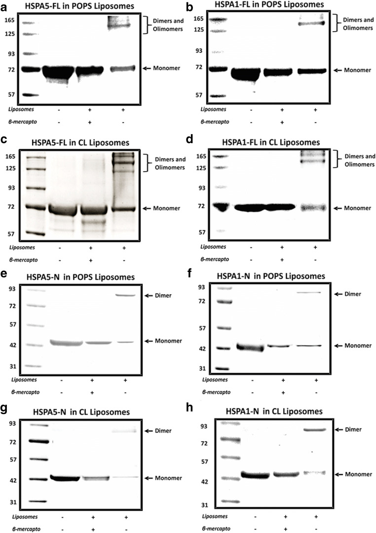
Fig. 7.
Aggregation profile of HSPA5 and HSPA1 and the respective N-terminal end domain after incorporation into liposomes. HSPA5-FL and HSPA5-N or HSPA1-FL and HSPA1-N and HSPA1-N were incorporated into POPS (a, b, e, f) or CL (c ,d ,g ,h) liposomes in 50 mM Tris-HCl buffer (pH 7.5) for 30 min at 25 °C. The mixture was centrifuged at 100,000×g for 1 h at 4 °C. The pellet was resuspended (300 μL) in 100 mM Na2CO3 buffer (pH 11.5) and centrifuged at 100,000×g for 1 h at 4 °C. The pellet was solubilized in sample buffer containing or not 10 mM βM, and the proteins were resolved by LDS-PAGE and visualized by staining with Coomassie Brilliant Blue R-250. The presence of dimers and oligomers is indicated.

