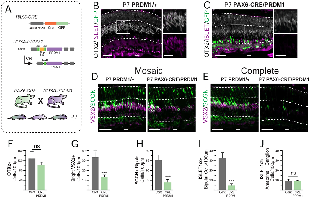
Figure 2. Constitutive PRDM1 expression driven by PAX6-CRE prevents bipolar cell formation at P7.

A) Schematic of transgenic mice utilized. B-C) P7 PRDM1/+ control compared to PAX6-CRE/PRDM1 retinas stained for ISLET1/2+ (purple), OTX2 (white), and GFP (green). Stain of VSX2 (purple) and SCGN (green) with D) mosaic loss and E) complete loss of bipolars in the peripheral retina. Note that SCGN marks some photoreceptors at this stage. F) The total number of OTX2+ cells (bright and faint) is not significantly decreased. G-I) Quantification of bipolar cell markers. There are fewer G) bright VSX2+ cells, H) SCGN+ bipolars, and I) ISLET1/2+ bipolars in in PAX6-CRE/PRDM1 retinas compared to controls. J) There is no difference in the number of ISLET1/2+ amacrines or ganglion cells between conditions. A total of 4,260 cells were quantified from 43 images. Statistics calculated based on number of mice (N), Cont N=4, PAX6-CRE/PRDM1 N=5. Error bars=standard deviation. ns=not significant, ***p<0.001. bars=50μm, inset bars=25μm, INL=Inner Nuclear Layer, ONL=Outer Nuclear Layer.
