FIGURE 5.
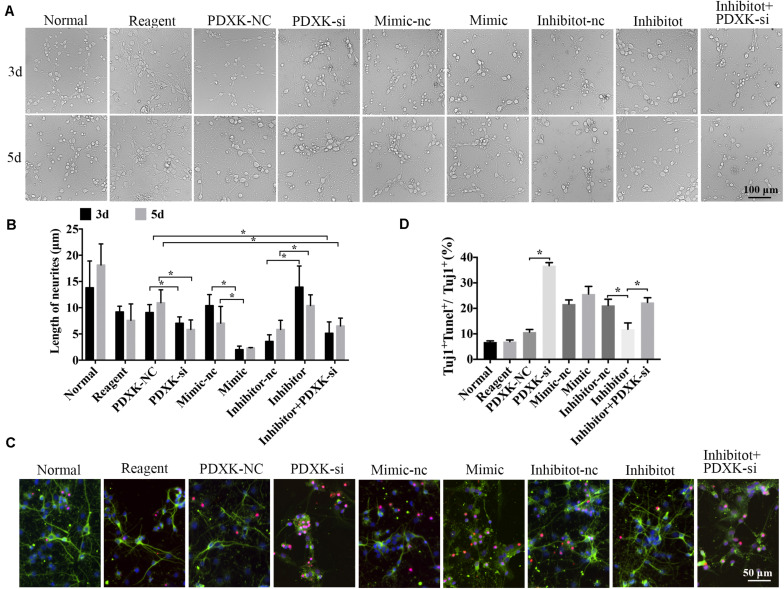
The role of miR-339 on neurite growth. (A) The cell physiological status under different situation in normal, reagent, NC, mimic-NC, inhibitor-NC, mimic, inhibitor, PDXK-si, and inhibitor + PDXK-si groups on 3 and 5 days after transfection. Scale bar = 100 μm. (B) Quantitative results of the length of neurites in normal, reagent, NC, mimic-NC, inhibitor-NC, mimic, inhibitor, PDXK-si, inhibitor + PDXK-si groups at 3 and 5 days post-transfection. (C) Evaluation of apoptotic cortical neurons by Tuj1 immunofluorescent staining and TUNEL staining, presented by Tuj1 + Tunel + /Tuj1 + (%) in normal, reagent, NC, mimic-NC, inhibitor-NC, mimic, inhibitor, PDXK-si, inhibitor + PDXK-si groups. DAPI counterstaining (blue) shows nuclei of intact cells, green fluorescence represented Tuj1 +, and red fluorescence represented apoptosis. Scale bar = 50 μm. (D) Quantitative analysis for neuronal apoptosis, presented by Tuj1 + Tunel + /Tuj1 + (%) in normal, reagent, NC, mimic-NC, inhibitor-NC, mimic, inhibitor, and PDXK-si, inhibitor + PDXK-si groups. Data were exhibited as mean ± SD; *P < 0.05. PDXK, pyridoxal kinase; NC, negative control; PDXK-si, PDXK interference; TUNEL, terminal deoxyribonucleotidyl transferase-mediated dUTP-digoxigenin nick-end labeling; DAPI, 4,6-diamidino-2-phenylindole.

