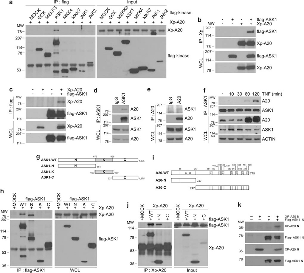
Figure 6.
A20 physically interacts with MAP3K, ASK1 in the JNK signaling cascade. (a) HEK293 cells were co-transfected with flag-tagged kinases (GCK, MEKK3, ASK1, MKK4, MKK7, JNK1, or JNK2) and Xp-tagged A20 expression plasmids. After immunoprecipitation with anti-flag antibody, co-immunoprecipitated proteins were detected by immunoblotting with anti-Xp antibody (top row). Expression levels of the transfected proteins in the immunoprecipitates and total cell lysates were analyzed by immunoblotting with anti-flag antibody (bottom row). (b) HEK293 cells were co-transfected with flag-tagged ASK1 and Xp-tagged A20 plasmids, as indicated. After immunoprecipitation with anti-Xp antibody, co-immunoprecipitated ASK1 was detected by immunoblotting with anti-flag antibody. The efficiency of immunoprecipitants and expression levels of the transfected proteins were analyzed by immunoblotting with the indicated antibodies. (c) Similar experiments as described in (b), with IP using anti-flag-antibody. (d) HEK293 cell extracts were immunoprecipitated with antibodies against ASK1 or normal IgG, and the resulting immunoprecipitates were analyzed by immunoblotting with anti-ASK1 and anti-A20 antibodies. (e) Similar experiments as described in (d), with IP using anti-A20 antibody. (f) HeLa cells were treated with TNF (15 ng/ml) for indicated times. After immunoprecipitation with anti-ASK1 antibody, co-immunoprecipitated A20 was detected by immunoblotting with anti-A20 antibody (top row). Expression levels of A20 and ASK1 in the total lysates were analyzed by immunoblotting with the indicated antibodies. (g, i) Schematic diagram of ASK1 and A20 domains for their expression constructs (see the text for details). (h, j, k) Various truncated forms of ASK1 and A20 were transfected in HEK293 cells. A20-ASK1 association was determined by immunoprecipitation and followed by immunoblotting with the indicated antibodies (top row). The efficiency of immunoprecipitants (h, j, bottom, left) and expression levels of the transfected proteins in the total lysates (h, j, bottom, right) were analyzed by immunoblotting with anti-flag and anti-Xp antibodies, respectively

