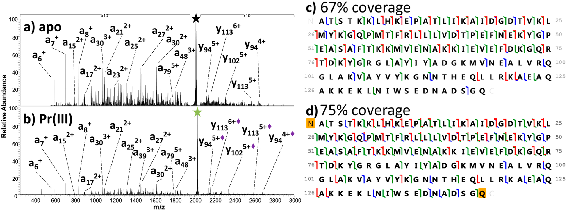
Figure 2.

UVPD of (a) apo SNase (8+) and (b) Pr(III)-bound SNase (8+) with some fragment ions labeled. Holo fragment ions (i.e., retaining the metal ion) are denoted with a diamond in the label. (c, d) Sequence coverage maps. Backbone cleavages that result in a/x (green), b/y (blue), and c/z• (red) fragment ions are indicated by the color-coded flags.
